Airway & pulmonary evidence · checked 2026-11-11
Airway care and adult lung function in STD and SCD: from infancy to adulthood.
Families reading older papers on Jarcho-Levin syndrome encounter frightening infant mortality figures next to stories of adults working, traveling, and raising children. Three clinical cohorts published between 2024 and 2026 finally connect both halves of that story: why the first eighteen months hinge on central airway geometry, and how survivors live full adult lives with 22% predicted vital capacity.
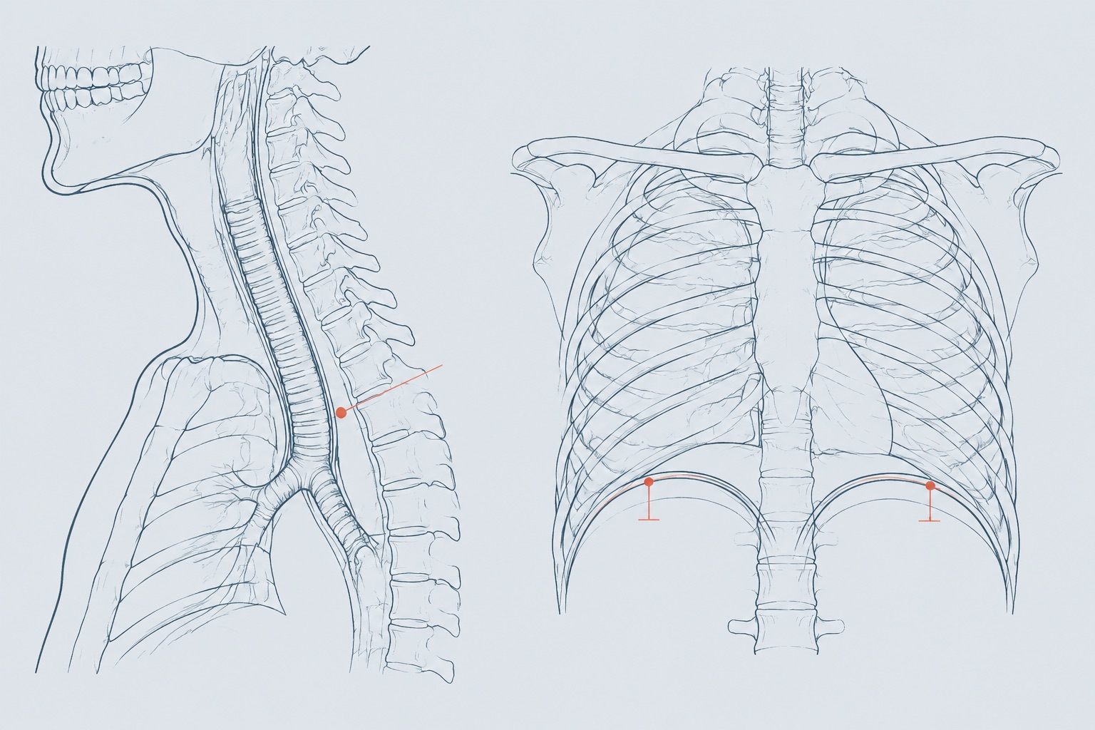
Short answer: In spondylothoracic dysostosis (STD) and severe spondylocostal dysostosis (SCD), respiratory risk peaks in infancy from shortened cervical airways, tracheal narrowing, and pneumonia. Patients who clear infancy adapt remarkably well: untreated STD adults average 22% predicted vital capacity yet report strong daily quality-of-life scores. JBJS 2024; J Surg Case Rep 2026; Radiology 2026
Why the infant airway is a cervical and mediastinal geometry problem, not just small lungs.
When clinicians explain the conditions once grouped under Jarcho-Levin syndrome, most diagrams focus on the ribs. In spondylothoracic dysostosis (STD), bilateral costovertebral rib fusions create a compact, symmetric chest cage; in spondylocostal dysostosis (SCD), asymmetric rib fusions and hemivertebrae shorten and twist the trunk. Yet for families in the neonatal intensive care unit (NICU), the most urgent crises often happen centimeters above the lungs, inside a shortened cervical airway and a crowded upper mediastinum.
In June 2026, pediatric surgeons in Ponce, Puerto Rico published a detailed technical case series on airway management in severe JLS (Journal of Surgical Case Reports 2026 airway series, DOI 10.1093/jscr/rjag405, PMID 42338708). Because Puerto Rico carries a well-documented founder prevalence of MESP2-related STD, where historical respiratory mortality reached up to 44% from pneumonia and restrictive physiology, the Ponce surgical team examined what actually happens when ventilated infants fail repeated extubation attempts during their first year of life.
Their report documents two structural bottlenecks that chest wall expansion devices such as VEPTR cannot solve on their own. First, fused or segmented cervical vertebrae produce an extremely short anterior neck with limited extension and a short subglottic-to-carina distance. In both ventilated infants, that foreshortened tracheal runway caused repeated accidental self-extubations whenever the baby moved or secretions built up. Second, the trachea sits low behind the sternal manubrium inside a crowded mediastinum, making standard off-the-shelf pediatric tracheostomy cannulas either slip out of the stoma or abut the carina.
To stabilize the airway safely, both infants required preoperative cross-sectional neck and chest imaging to measure exact tracheal length, angulation, and great-vessel relationships before surgery. In the first infant, born at 30 weeks of gestation with severe ventilatory dependence, dissection 1 cm above the sternal notch could not expose the low-lying trachea until the surgeons performed a superior partial manubriectomy using a Lebske knife and placed a custom wire-reinforced silicone tracheostomy tube measuring 4.5 F by 46 mm. In the second infant, diagnosed prenatally at 20 weeks and reliant on non-invasive ventilation before recurrent admissions forced tracheostomy at 15 months of age, imaging guided placement of a custom wire-reinforced silicone Bivona tube measuring 3.5 F by 30 mm with thyroid isthmus division and tracheal stay sutures.
Equally important for parents weighing intensive care decisions is the honest timing lesson from that series. Although both customized tracheostomies were completed with zero intraoperative complications, both infants had already developed severe downstream sequelae of chronic hypoventilation and recurrent infection before surgical referral: refractory multidrug-resistant pneumonia in the first child, and echocardiographically confirmed pulmonary artery dilation with moderate pulmonary hypertension requiring sildenafil by three months of age in the second. Both children died months later (at 13 and 18 months of age) from progressive cardiorespiratory failure. Once fixed pulmonary vascular remodeling and parenchymal damage take hold, securing the airway late cannot reverse them. Early multidisciplinary airway evaluation must happen before recurrent pneumonia and pulmonary hypertension become established.
Anesthesia, endotracheal intubation, and the hidden trap of thoracic inlet narrowing.
Even children on the milder end of the SCD or STD spectrum frequently need general anesthesia for inguinal hernia repair, sedated MRI scans, ear tubes, or orthopedic procedures. Every anesthetic induction in a child or adult with costovertebral segmentation anomalies requires difficult-airway preparation rather than routine direct laryngoscopy. Cervical block vertebrae restrict neck extension into the classic sniffing position, high diaphragmatic resting pressure accelerates oxygen desaturation during apnea, and neuraxial landmarks are distorted by lumbar hemivertebrae, as highlighted in obstetric and perioperative case audits (BMJ Case Reports 2020 STD anesthesia report, DOI 10.1136/bcr-2019-232964; A&A Practice 2024 SCD perioperative report, DOI 10.1213/xaa.0000000000001881). Maintaining spontaneous ventilation until the glottis is secured with videolaryngoscopy or a flexible fiberoptic bronchoscope is a core safety rule.
A 2026 surgical report from Nanjing Drum Tower Hospital uncovered an even subtler airway hazard tied directly to growth-friendly spine distraction (BMC Surgery 2026 thoracic inlet stenosis study, DOI 10.1186/s12893-026-03881-5, PMID 42226213). Many children with congenital thoracolumbar kyphoscoliosis develop compensatory upper thoracic hyperlordosis, sometimes called a straight-back profile, which quietly narrows the anteroposterior gap between the upper thoracic vertebral bodies posteriorly and the sternal manubrium anteriorly.
In that 2026 report, a 6-year-old child tolerated initial dual growing-rod implantation without respiratory trouble. One year later, during the first scheduled rod distraction, lengthening the posterior spinal construct flattened upper thoracic alignment further, pinching the trachea inside the thoracic inlet. She suffered acute airway spasm and hypoxemia during intubation and could not be extubated after surgery. Bronchoscopy and computed tomography confirmed critical extrinsic tracheal compression at the thoracic inlet. Loosening the rods alone failed to reopen the airway; definitive relief required multilevel posterior column osteotomies (PCOs) paired with kyphotic-contoured rods to restore natural upper thoracic kyphosis, allowing extubation on postoperative day two and symptom-free breathing at one-year follow-up. For families already navigating the six-month lengthening calendar, checking sagittal thoracic-inlet clearance on lateral imaging before distraction is a practical safeguard.
Crossing out of infancy: the 22% vital capacity paradox in untreated STD adults and 4D MRI diaphragm mechanics.
Parents searching late at night often face an agonizing contradiction: if an infant survives the early months without chest wall expansion surgery, does lung function inevitably deteriorate in adolescence or adulthood? For decades, textbooks assumed the answer was yes, treating a radiographic diagnosis of STD as an automatic surgical mandate. Two landmark Puerto Rican natural-history cohorts led by Norman Ramírez, Alberto Santiago Cornier, and Ernesto Rosado, alongside a 149-patient dynamic MRI investigation published in 2026, tell a far more nuanced story.
First, separating SCD from STD clarifies early survival odds. In a prospective cohort of 13 children with spondylocostal dysostosis managed systematically with regular chest physiotherapy and selective surgery in only two refractory cases (Spine 2004 prospective SCD cohort, DOI 10.1097/01.brs.0000128761.72844.ab, PMID 15223937), survival at a mean 4.5-year follow-up was 100%, with respiratory infections declining steadily as airways matured. By contrast, in the classic 28-patient multicenter STD study (Journal of Bone and Joint Surgery 2007 STD natural history, DOI 10.2106/jbjs.f.01085, PMID 18056499), eight infants (29%) died during the neonatal period. Yet among the 20 STD survivors evaluated later in childhood, despite a posterior thoracic spine averaging just 24.2% of predicted normal length, CT lung volumes averaging 28% of expected values, mean forced vital capacity (FVC) of 27.9% predicted, and mean forced expiratory volume in one second (FEV1) of 29.5% predicted (FEV1/FVC ratio 0.92), children were clinically active without progressive congenital deformity.
In 2024, Ramírez, Rosado, Cornier, and colleagues answered what happens decades later by evaluating 11 skeletally mature, surgically untreated adults with genetically and radiographically confirmed STD aged 15 to 70 years, with a mean age of 32.3 years (Journal of Bone and Joint Surgery 2024 untreated adult STD study, DOI 10.2106/jbjs.23.00800, PMID 38127843). Across five men and six women, mean thoracic height measured only 16 cm (range 12 to 17 cm) and mean body mass index was 24.4 kg/m². Spirometry confirmed profound static restriction: mean FVC stood at 22% of predicted, mean FEV1 at 24% of predicted, and FEV1/FVC at 107% of predicted, indicating brisk, unobstructed airflow through a very small lung volume.
What startled orthopedic and pulmonary specialists was how those adults actually felt and functioned. Their mean Patient-Reported Outcomes Measurement Information System (PROMIS) dyspnea score was 40 ± 8 points (below the general-population reference mean of 50 for symptom burden), and their mean 24-Item Early Onset Scoliosis Questionnaire (EOSQ-24) total score reached 77.3 ± 18 points (range 43.9 to 93.2), outperforming published cohorts with neuromuscular scoliosis. Neither static FVC percentage nor thoracic height in centimeters predicted a person's daily dyspnea or quality-of-life score, leading the authors to conclude that an STD diagnosis alone should no longer be treated as an absolute indication for VEPTR expansion thoracoplasty.
How can an adult work and walk comfortably on 22% predicted vital capacity? A 2026 study of 149 pediatric TIS patients compared with 190 healthy controls using four-dimensional free-breathing dynamic MRI provides the biomechanical explanation (Radiology: Cardiothoracic Imaging 2026 4D dMRI diaphragm study, DOI 10.1148/ryct.250345, PMID 42312935). Unlike neuromuscular disorders where respiratory muscles weaken, children and adults with STD and SCD retain intact phrenic nerve drive and strong abdominal-diaphragmatic coupling. Quantifying three-dimensional curvature across 13 regions per hemidiaphragm, researchers found that central sagittal diaphragm curvature correlated directly with real-world ventilatory independence (correlation coefficient up to 0.392, P < 0.001), and when VEPTR surgery was performed in decompensated children, right anterior-lateral end-expiratory sagittal dome curvature rose from 6.3 ± 0.7 m⁻¹ to 8.2 ± 0.6 m⁻¹ (P = 0.02), approaching healthy control curvature (9.3 ± 2.6 m⁻¹). When the rib cage is fused, the diaphragm piston carries the life of the lung.
Genotype-to-airway comparison and the family multidisciplinary clinic checklist.
Because hospital emergency teams and pre-anesthesia clinics rarely see more than one or two patients with costovertebral dysostosis in a career, families benefit from carrying a clear summary of how their child's subtype behaves. You can pair the table and checklist below with our broader postnatal breathing and specialty care guide before scheduled appointments.
| Clinical & airway dimension | Spondylothoracic dysostosis (STD · MESP2) | Spondylocostal dysostosis (SCD · DLL3, LFNG, HES7, TBX6, RIPPLY2, DMRT2) |
|---|---|---|
| Cervical spine & upper airway geometry | Markedly shortened neck with cervical segmentation defects and a low-lying trachea behind the manubrium; short vocal-cord-to-carina distance raises accidental extubation risk in ventilated infants. | Variable cervical block vertebrae and rotational asymmetry; neck extension may still be restricted even when external neck length looks near-typical. |
| Anesthesia & intubation protocol | High difficult-airway risk plus rapid desaturation from low functional residual capacity; preserve spontaneous breathing and use videolaryngoscopy or fiberoptic bronchoscopy. | Preoperative cervical mobility exam mandatory; screen for compensatory upper thoracic lordosis ("straight back") that can narrow the thoracic inlet during spine distraction. |
| Infant respiratory risk window (0 to 18 months) | Primary life-threatening bottleneck (29% neonatal mortality in 2007 multicenter series; up to 44% historically in severe cohorts) driven by pneumonia, atelectasis, and secondary pulmonary hypertension. | Mild-to-moderate neonatal tachypnea in most subtypes (100% 4.5-year survival in the 13-patient 2004 prospective physiotherapy series), though severe biallelic forms occur. |
| Tracheostomy technical considerations | Evaluate early if repeated extubation failures occur, prior to fixed pulmonary hypertension; requires neck/chest CT sizing, custom wire-reinforced silicone tubes, and occasional partial manubriectomy. | Rarely required long-term; if prolonged ventilation is needed in severe phenotypes, CT measurement of tracheal length and thoracic-inlet caliber prevents cannula malposition. |
| Childhood & adult spirometry vs daily life | Severe static restriction persists (27.9% predicted FVC in childhood; 22% predicted FVC and 16 cm thoracic height in untreated adults aged 15 to 70), yet FEV1/FVC stays normal (107% pred) and EOSQ-24 averages 77.3/100. | Mild-to-moderate restrictive pattern proportional to thoracic scoliosis and rib fusion asymmetry; most individuals reach independent adulthood without home ventilatory support. |
Six-point family checklist for pulmonology, anesthesia, and surgical visits
- Carry an anesthesia airway card before any sedated scan or minor surgery: note cervical vertebral fusions, request pediatric anesthesiologists experienced with videolaryngoscopy or fiberoptic intubation, and avoid unnecessary neuromuscular paralysis until airway view is confirmed.
- Screen the right heart early when an infant needs prolonged oxygen or CPAP: ask for an echocardiogram in early infancy (around 3 months or sooner if oxygen needs climb) to detect pulmonary artery dilation or early pulmonary hypertension while it is still treatable.
- Check the thoracic inlet in profile before spinal or rib distraction: if your child has a flat upper back (thoracic hypokyphosis or lordosis) and is scheduled for growing-rod or VEPTR lengthening, ask the surgeon to review anteroposterior tracheal space behind the manubrium.
- Protect diaphragm excursion from below: because the diaphragm dome generates nearly all tidal volume in a stiff rib cage, treat gastroesophageal reflux, constipation, abdominal distension, and nutritional depletion aggressively so upward abdominal pressure does not splint the diaphragm.
- Pair airway clearance with a rapid respiratory-infection plan: establish daily chest physiotherapy techniques when secretions thicken, keep RSV and influenza immunizations current, and agree on an early-assessment threshold before minor viral bronchiolitis turns into lobar atelectasis.
- Judge adult and adolescent surgical candidacy by function, not FVC% alone: in older children and adults with STD, combine sleep capnography/oximetry, endurance, infection frequency, and dynamic diaphragm imaging rather than treating a low static spirometry percentage as an automatic crisis.
Questions families bring to the airway, anesthesia, and pulmonology team.
Why can standard pediatric tracheostomy or breathing tubes slip out or obstruct in infants with STD or severe SCD?
Shortened cervical vertebrae, limited neck extension, a high sternal manubrium, and a short distance from the vocal cords to the carina leave very little tracheal length. In a 2026 Journal of Surgical Case Reports series from Puerto Rico (PMID 42338708), standard tubes repeatedly dislodged until cross-sectional imaging guided custom wire-reinforced silicone tubes (4.5 F by 46 mm and 3.5 F by 30 mm) and, in one infant, a superior partial manubriectomy.
Can spinal growing-rod distraction narrow the windpipe even while straightening the spine?
Yes, in children who have compensatory upper thoracic lordosis (a flat or straight upper back). A 2026 BMC Surgery clinical report (PMID 42226213) documented critical extrinsic tracheal stenosis at the thoracic inlet triggered during growing-rod distraction, causing intraoperative bronchospasm and failed extubation until posterior column osteotomies and kyphotic rod contouring restored space behind the manubrium.
Can a child with spondylothoracic dysostosis survive to adulthood without VEPTR surgery, and what do adult lung tests show?
Yes, once a child clears the high-risk infant window. A 2024 Journal of Bone and Joint Surgery follow-up of 11 skeletally mature, surgically untreated adults with STD aged 15 to 70 years (mean 32.3 years, PMID 38127843) recorded a mean forced vital capacity (FVC) of 22% predicted and thoracic height of 16 cm, yet mean EOSQ-24 quality-of-life score was 77.3 points and PROMIS dyspnea was 40 points, showing strong everyday tolerance driven by intact diaphragm mechanics.
Sources. Zapata-Valentin M, Soto PM, Nater LM, Urban JC, Justiniano VO. Surgical management of the airway in Jarcho-Levin syndrome: a case series (Journal of Surgical Case Reports 2026, DOI 10.1093/jscr/rjag405, PMID 42338708). Sun S, Li S, Liu Z, Shi B, Qiu Y, Zhu Z, Mao S. Iatrogenic thoracic inlet narrowing during growing rod distraction causing critical tracheal stenosis (BMC Surgery 2026, DOI 10.1186/s12893-026-03881-5, PMID 42226213). Dindarian S, Shaghaghi S, Hosseini M et al. Free-breathing Dynamic MRI Analysis of Three-dimensional Diaphragm Curvatures in Pediatric Patients with Thoracic Insufficiency Syndrome (Radiology: Cardiothoracic Imaging 2026, DOI 10.1148/ryct.250345, PMID 42312935). Ramírez N, Monroig-Rivera C, De Jesús-Rojas W, Rosado E, Cornier AS et al. Long-Term Follow-up of Untreated Adult Patients with Spondylothoracic Dysostosis (Journal of Bone and Joint Surgery 2024, DOI 10.2106/jbjs.23.00800, PMID 38127843). Ramírez N, Cornier AS, Campbell RM Jr et al. Natural history of thoracic insufficiency syndrome: a spondylothoracic dysplasia perspective (Journal of Bone and Joint Surgery 2007, DOI 10.2106/jbjs.f.01085, PMID 18056499). Teli M, Hosalkar H, Gill I et al. Spondylocostal dysostosis: thirteen new cases treated by conservative and surgical means (Spine 2004, DOI 10.1097/01.brs.0000128761.72844.ab, PMID 15223937). Dolinski L et al. Anaesthetic management of a parturient with spondylothoracic dysostosis (BMJ Case Reports 2020, DOI 10.1136/bcr-2019-232964, PMID 31980478). J Surg Case Rep 2026; BMC Surg 2026; Radiol Cardiothorac Imaging 2026; JBJS 2024; JBJS 2007; Spine 2004