Surgical evidence · checked 2026-10-22
The VEPTR lengthening decade: what six-month expansions actually do for a fused chest.
Surgeons describe Vertical Expandable Prosthetic Titanium Rib (VEPTR) surgery as growth-sparing. For families, it is a ten-year calendar of six-month hospital returns, scar care, and room made millimeter by millimeter while alveoli are still multiplying.
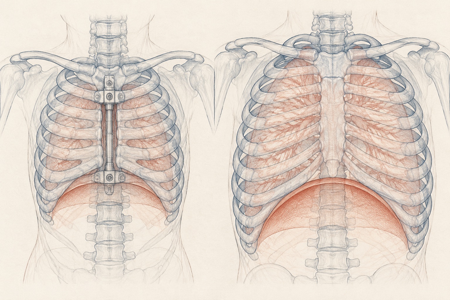
Short answer: Vertical Expandable Prosthetic Titanium Rib (VEPTR) surgery treats thoracic insufficiency syndrome by separating fused or constricted ribs and anchoring a sliding titanium cradle that is lengthened roughly every six months. Dynamic 4D MRI shows VEPTR increases chest wall and diaphragm tidal volume by a mean of 70% even when standing spine X-ray angles barely change, while carrying a 13% per-procedure skin or hardware complication risk across childhood. J Pediatr Orthop 2020; Eur Spine J 2010; Spine 2005
Why a flat X-ray misses the real gain inside the chest.
Parents who spend years in pediatric orthopedic waiting rooms learn to ask for the Cobb angle first. In idiopathic scoliosis, that single degree measurement on a standing radiograph tells most of the story. In spondylothoracic dysostosis (STD) and severe spondylocostal dysostosis (SCD), it frequently misleads. A chest with posterior rib fusions or a short, stiff thoracic spine is a three-dimensional piston problem: the question is not how straight the spine looks on a wall film, but how many milliliters of air the rib cage and diaphragm can move with each breath.
In 2020, dynamic magnetic resonance imaging finally quantified what families and pulmonologists had observed at the bedside (Journal of Pediatric Orthopedics, DOI 10.1097/BPO.0000000000001258). Researchers followed 25 consecutive patients with thoracic insufficiency syndrome (7 congenital, 14 neuromuscular, 4 other; preoperative thoracic Cobb angles ranging from 0° to 136°) before and after VEPTR implantation using quantitative 4D dynamic MRI. Across the cohort, postoperative chest wall and diaphragmatic tidal volume components rose by a mean of 70%, with individual gains reaching 533%.
Just as important for clinic conversations was what did not predict breathing improvement: static spine curvature angles on radiographs showed no significant correlation with tidal volume gains. Instead, thoracolumbar deformity restricted downward diaphragm excursion far more severely than isolated main thoracic curves. Freeing the lower rib cage and giving the diaphragm dome room to descend did work that no two-dimensional Cobb angle could capture.
What the six-month lengthening calendar actually looks like.
In the landmark prospective trial by Robert Campbell Jr. and Anna Kathrin Hell-Vocke (Spine 2005, DOI 10.1097/01.brs.0000175194.31986.2f), 31 children with fused ribs and thoracic insufficiency syndrome underwent opening-wedge expansion thoracostomy and VEPTR placement after early spinal arthrodesis was rejected because fusing a toddler's spine permanently halts thoracic growth. In 30 of those 31 children, deformity was controlled and the thoracic spine continued to grow at near-normal rates during follow-up.
The honest cost of keeping that growth open is repetition. Because a titanium sleeve does not grow on its own, a child returns to the operating theatre roughly every six months through childhood so the surgeon can open a small incision over the connector lock and distract the device by a few millimeters. A longitudinal cohort by Hasler, Mehrkens, and Hefti (European Spine Journal 2010, DOI 10.1007/s00586-009-1253-9) audited 187 consecutive VEPTR procedures across 23 early-onset patients (mean age 6.5 years at implantation, minimum two-year follow-up): 23 index implantations, 149 scheduled six-month lengthenings (an average of 6.5 lengthenings per child in that window), and 15 unplanned procedures.
Across all 187 surgeries, the authors recorded 23 complications, or 0.13 per surgical procedure (affecting 9 of the 23 children). Ten were skin sloughs over prominent hardware in thin children with scarce subcutaneous tissue; six were infections; five were device dislocations or rib-hook migrations through soft pediatric bone; and two were rod breakages. As the 2022 state-of-the-art review in Paediatric Respiratory Reviews (DOI 10.1016/j.prrv.2022.02.003) emphasizes, nutrition before implantation, soft-tissue flap coverage, and delaying definitive spinal fusion until skeletal maturity are what allow those lengthening years to translate into adult lung reserve.
How chest architecture shapes the surgical decision in STD vs SCD.
Families reading hospital brochures often see VEPTR, magnetically controlled growing rods (MCGR), and traditional dual growing rods grouped together. In practice, the anatomy of the rib cage dictates which device can anchor safely and whether surgery is needed at all. You can compare this directly with our broader postnatal care and breathing support guide.
| Clinical dimension | Spondylothoracic dysostosis (STD · typically MESP2) | Spondylocostal dysostosis (SCD · DLL3, LFNG, HES7, TBX6) |
|---|---|---|
| Rib and spine geometry | Symmetric posterior fusion of ribs at the costovertebral junctions ("crab-like" or fan-like thorax); short, rigid thoracic column with minimal sideways scoliosis. | Asymmetric rib fusions, absent ribs, and multiple hemivertebrae or block vertebrae; higher risk of progressive thoracic or thoracolumbar scoliosis. |
| Primary breathing bottleneck | Severe vertical restriction of the chest cage and high diaphragmatic work of breathing in infancy. | Asymmetric hemithorax restriction; thoracolumbar curves can tether and flatten diaphragm excursion on the concave side. |
| Typical surgical strategy when TIS develops | Bilateral or staged rib-to-rib / rib-to-pelvis VEPTR cradles to lengthen vertical thoracic height and lower the diaphragm dome. | Opening-wedge thoracostomy with unilateral or bilateral VEPTR when ribs are fused, or growth-friendly spinal constructs when deformity is primarily spinal. |
| What happens at skeletal maturity | Lengthenings stop once thoracic growth plateaus; devices are retained, revised, or selectively converted depending on chest wall stability. | Transition from six-month lengthenings to definitive spinal fusion or final construct stabilization once alveolar and trunk growth are complete. |
Questions families bring to the thoracic insufficiency clinic.
Why does a standing X-ray Cobb angle fail to show whether VEPTR helped a child breathe?
A flat spine X-ray only measures static bone curvature, not chest wall motion or diaphragm excursion. In the 25-patient dynamic 4D MRI study (Journal of Pediatric Orthopedics 2020), VEPTR surgery increased chest wall and diaphragm tidal volume components by a mean of 70% (up to 533%), and static Cobb angle did not correlate with dynamic breathing volume gains.
How often are VEPTR rods lengthened and what is the real complication rate over childhood?
Expansion procedures are typically scheduled every 6 months during active thoracic growth. In the 187-surgery European Spine Journal cohort of 23 early-onset patients (mean 6.5 lengthenings per child, DOI 10.1007/s00586-009-1253-9), there were 23 total complications (0.13 per surgery): 10 skin sloughs over prominent hardware, 6 infections, 5 device migrations or dislocations, and 2 rod breakages.
Do children with spondylothoracic dysostosis (STD) and spondylocostal dysostosis (SCD) need the same chest surgery?
Not always. In STD (MESP2), ribs fuse symmetrically at the spine in a fan or crab-like configuration that restricts vertical chest height, whereas in SCD asymmetric rib fusions and hemivertebrae often drive progressive thoracic or thoracolumbar scoliosis. Surgical teams evaluate dynamic breathing mechanics, oxygen or CPAP dependence, and rib fusion anatomy before recommending opening-wedge thoracostomy with VEPTR.
Sources. Tong Y, Udupa JK, McDonough JM, Wileyto EP, Capraro A, Wu C, Ho S, Galagedera N, Talwar D, Mayer OH, Torigian DA, Campbell RM Jr. Quantitative Dynamic MRI (dMRI) Volumetric Analysis of Pediatric Patients with Thoracic Insufficiency Syndrome. Journal of Pediatric Orthopedics, 2020. DOI 10.1097/BPO.0000000000001258 (PMID 32132448). Campbell RM Jr, Hell-Vocke AK. Growth of the thoracic spine in congenital scoliosis after expansion thoracoplasty. Spine, 2005. DOI 10.1097/01.brs.0000175194.31986.2f (PMID 16138067). Hasler CC, Mehrkens A, Hefti F. Efficacy and safety of VEPTR instrumentation for progressive spine deformities in young children without rib fusions. European Spine Journal, 2010. DOI 10.1007/s00586-009-1253-9 (PMID 20041270). Reddy S et al. Thoracic insufficiency syndrome: an overview. Paediatric Respiratory Reviews, 2022. DOI 10.1016/j.prrv.2022.02.003 (PMID 35339395). J Pediatr Orthop 2020; Spine 2005; Eur Spine J 2010; Paediatr Respir Rev 2022